Drawing regions-of-interest (ROIs)
Manual delineation of ROIs is often considered as gold standard although it is a time-consuming and error-prone task with considerable inter- and intra-operator variability.
Software for defining ROIs manually in TPC
Currently ROIs (or VOIs = volumes of interest) can be drawn on MR and parametric or sum PET images using:
- CarimasTM relase 1.x for myocardium and upcoming 2.0 for all target organs.
- Imadeus on PC/Windows. Images must be in Analyze format. ROI format is not yet supported by other programs, although Carimas 2.x can read and convert Imadeus ROI format. Time-activity curve (TAC) file format is fully supported.
- Vinci on PC/Windows. Images can be in several formats, including ECAT 7.x and Interfile. If TACs are saved in CPT format, they can be converted to our standard DFT format using program cpt2dft.
- YaIT on all Solaris workstations. Images can be in ECAT 7.x or 6.3 format. ROIs are saved and TACs calculated using program img2dft.
- Xeleris workstation; TAC files calculated in Xeleris can be converted to DFT files.
- Amide: TAC data in *.tsv files can be converted to DFT format: tsv2dft
In addition, ROIs can be drawn using GE Display on GE Advance images, but TACs produced can not be extracted from the database and used in further calculations, except the ones provided in the Display program.
Calculation of regional TACs
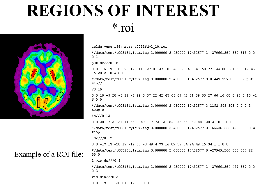
ROI files
ECAT ROI files
Format of ECAT ROI files; see also PET C-library, roi.c and roi.h
Working with ROI files:
- Listing ROI file contents:
roilist - Splitting ROI file into parts:
roisplit - Calculating ROI TACs:
img2dftin Sun/Solaris and PC/Windows, or img2kbq on Sun/Solaris - Seeing ROI placement by setting
blank the outside areas in images:
eroi2img - Masking high-activity image
regions by setting pixel values inside ROIs to zero:
eroi2img - Extracting TACs of separate
pixels from dynamic image:
roipxl - Extracting pixel values inside
ROI(s):
imgpext(replacesextrimg) - Extracting one image line:
lor2dft - Find out the size of ROIs
Imadeus ROI files
Working with Imadeus ROIs.
Automatic ROI delineation
An automated anatomy-based region-of-interest (ROI) analysis can be performed using standardized ROIs defined on magnetic resonance template image representing brain anatomy in accordance with the Montreal Neurologic Institute (MNI) space database (Nagano et al., 2000; Brück et al., 2005).
