[15O]CO - Measurement of blood volume
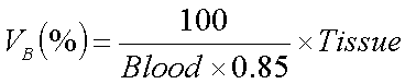
Blood volume (VB) in the brain has been measured with PET using carbon monoxide as a tracer labeled with either C-11 or O-15 (Grubb et al., 1978; Martin et al., 1987). The model is very simple: inhaled carbon monoxide is assumed to bind and stay bound to hemoglobin in blood. After the "labeled" red blood cells are distributed evenly in the vasculature, the concentrations of radioactivity in tissue and in blood are measured. To get an estimate of the volume fraction of blood in tissue, the tissue radioactivity concentration is divided by concentration in blood.
For precise quantitation, the difference between hematocrit values in tissue vasculature and in large veins where blood sampling is done, must also be accounted for. The ratio of hematocrits in the brain is usually assumed to be 0.85.
The blood volume as percentage of tissue volume can be calculated from equation
In Turku PET Centre, the unit of radioactivity concentration is always
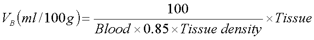
kBq/ml. Therefore, if VB is needed per mass of tissue
(ml blood/100 g tissue) the result must be divided by density of the tissue
(density of the brain is 1.05 g/ml):
If VB is needed as a fraction (ml blood/ml tissue) instead of percentage, use '1' as dividend in the first equation instead of '100'.
Unless otherwise stated, all programs mentioned below are available on Solaris workstations and PC/Windows.
Brain
[15O]CO PET study
Labeled carbon monoxide is inhaled for about 2 min (Martin et al., 1987). After the inhalation, two minutes are waited before the beginning of a single 4-min scan. During the sacnning, three whole blood samples are taken; samples can be either arterial or venous (Martin et al, 1987).
Calculation of VB image
- Use a pocket calculator, or preferably a spreadsheet program, to calculate a
coefficient with which the PET image will be multiplied with.
- Calculate an average of the measured three values of blood radioactivity. The blood datafile is in ASCII format and can be viewed with any word processor or listed on-screen with command 'type' (Windows/MS-DOS) or 'more' (Unix/Linux) followed by the filename
- Multiply the blood average by 0.85
- Divide 100 by this product value; i.e. 100/(blood*0.85)
- If VB is needed in unit ml blood/100 g tissue (i.e. per mass, not per volume), the value must be further divided by density of the tissue, e.g. 1.05
- Multiply the PET image with this value using program
ecatcalc:
ecatcalc PETimage x Value VBimage