[Turku PET Centre] [PET data analysis] [Main page] [Previous page] [Next page]
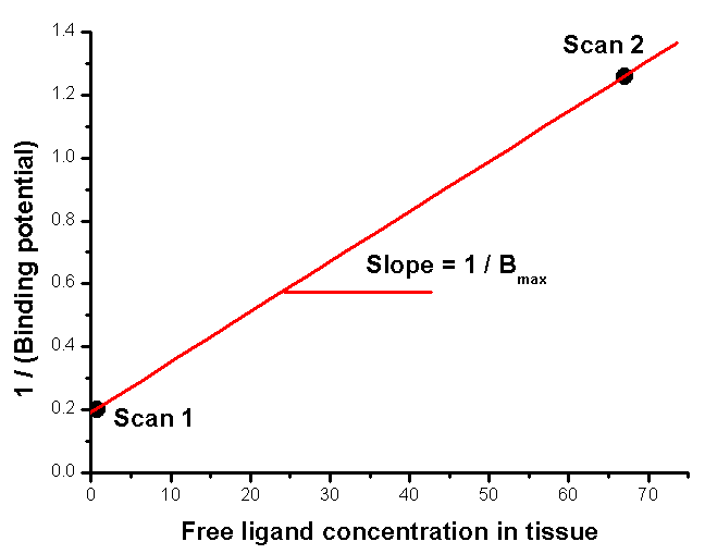
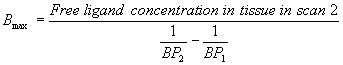
Note: here is newer version of this page.Calculation of "binding potential", k3/k4, at two (or more) ligand concentrations makes it possible to determine the regional Bmax and KD of the receptor ligand. If binding is irreversible during PET scan, also k3 can applied (Wong et al 1986, 1997).
When k3/k4, or an estimate of it, is measured under two different ligand concentrations, the data can be plotted against the concentration of free ligand (tracer ligand + possible drug). The high ligand concentration can be achieved either by injecting the labeled ligand with large amount of cold ligand (low specific activity), or by giving another inhibitor, e.g. an oral dose of a drug compound that binds to the same receptor.
The concentration of the free ligand in the tissue can be estimated from the radioactivity concentration in a reference region (receptor free tissue), divided by specific activity, when the labeled ligand and cold ligand (inhibitor) are injected together (Farde et al 1986, 1987). If the first PET study is performed with tracer dose, the concentration of the free ligand can be assumed to be zero. In that case,
If the second PET scan is performed with tracer dose of PET ligand and high
dose of another inhibitor, the concentration of free ligand in scan 2 can be
estimated based on the plasma concentration of the drug and the free
tissue-to-plasma ratio of the drug. This ratio can be estimated from the same
PET study as the distribution volume (Vd) of the drug in a reference
region (arterial plasma sampling is required), or if a receptor free region does
not exist, as the distribution volume of an enantiomer of the drug that does not
bind to the receptor. Estimates of the ratio can also be measured in ex vivo or
in vitro human and animal studies (Wong et al 1986, 1997). When the free ligand
concentration is calculated as Vd * ligand concentration in plasma,
it must be noticed that both measures either have to be corrected for binding to
plasma proteins, or if binding of PET tracer and the drug is similar, both can
be left uncorrected.
Farde L, Hall H, Ehrin E, Sedvall G. Quantitative analysis of D2 dopamine receptor binding in the living human brain by PET. Science 1986; 231(4735): 258-261.
Farde L, Halldin C, Stone-Elander S, Sedvall G. PET analysis of human dopamine receptor subtypes using 11C-SCH 23390 and 11C-raclopride. Psychopharmacology (Berl). 1987; 92: 278-284.
Gjedde A, Wong DF. Modeling neuroreceptor binding of radioligands in vivo. In: Quantitative imaging, neuroreceptors, neurotransmitters, and enzymes, edited by JJ Frost and HN Wagner Jr, Raven Press, New York, 1990, 51-79.
Wong DF, Gjedde A, Wagner HN Jr. Quantification of neuroreceptors in the living human brain. I. Irreversible binding of ligands. J Cereb Blood FlowMetab 1986; 6: 137-146.
Wong DF, Young D, Wilson PD, Meltzer CC, Gjedde A. Quantification of neuroreceptors in the living human brain. III. D2-like dopamine receptors: theory, validation, and changes during normal aging. J Cereb Blood Flow Metab 1997; 17: 316-330.