[Turku PET Centre] [PET data analysis] [Main page] [Previous page] [Next page]
Positron emission tomography (PET) is an imaging method for quantitatively measuring biochemical and physiological processes in vivo by using radiopharmaceuticals and by measuring the annihilation radiation using a coincidence detecting technique.
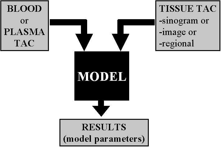
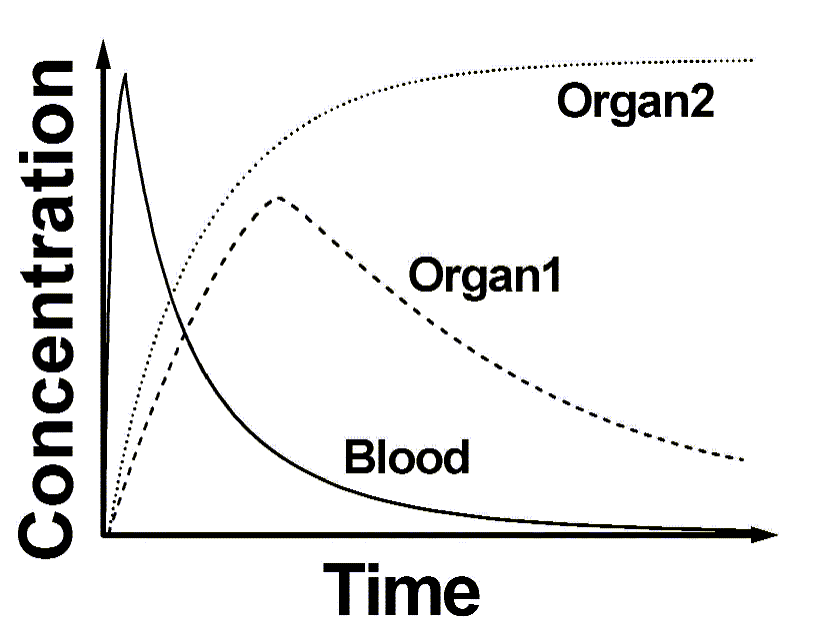
Physiologically active compounds are tagged with positron-emitting isotopes of oxygen, nitrogen, carbon or fluorine, while maintaining their biological properties. The labeled compounds are administered intravenously in tracer amounts (e.g., in nanomolar or picomolar quantities). Local rates of accumulation, fixation, or turnover of tracer in tissue are measured in absolute units of tissue concentrations from the cross-sectional PET images and represent quantitatively regional tracer tissue concentrations. From the tissue response to an arterial tracer input function, tracer kinetic models are developed that relate the kinetics of a tracer in tissue to the physiological process under study, allow description of the tissue kinetics in mathematic terms, and enable an operational equation to be formulated by which absolute rates of blood flow and metabolism can be calculated.
PET scanner can deliver quantitative images of radioactivity (Bq/voxel, or kBq/mL). The chemical identity of the labeled molecules is only known at the moment of injection. Due to metabolism in blood, in the tissue region of interest, and in other tissues, different labeled compounds are formed in the course of time. The relative amounts of different radioactive compounds in plasma can be measued from arterial blood samples as a function of time.