Image reconstruction
The process where actual PET image is formed from the raw data (sinograms) collected by the PET scanner.
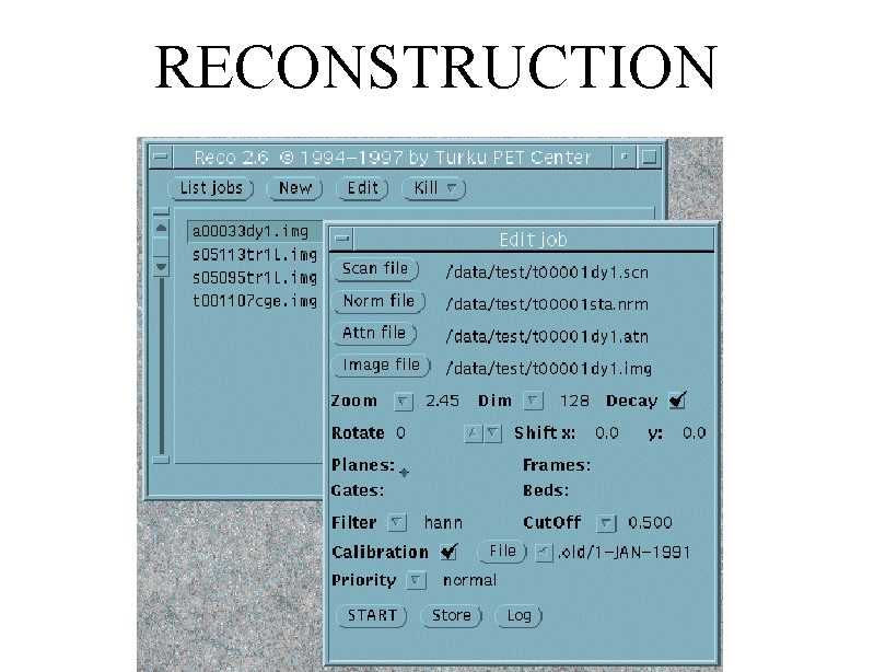
PET images are automatically reconstructed from the raw data by the personnel of PET centre, and images are stored in PETO. However, if data from our first scanner, ECAT 931, need to be reconstructed again, then that must be done by the researcher. Most of this page deals with reconstruction of ECAT 931 data.
Reconstruction of ECAT 931 images
Previously, GUI program for reconstruction of ECAT 931 and 2D GE Advance sinograms in CTI ECAT 6.3 format was found in the menus on SUN/Solaris platforms. This is no longer functional, but command-line versions of reconstruction and calibration correction programs can be used from SUN/Solaris Unix command-prompt.
Required data
For reconstruction you need obviously the sinograms (*.scn), but you also need the attenuation and normalization files (*.atn and *.nrm).
After reconstruction, images need to be calibrated to units kBq/mL, and for that you need appropriate calibration file (see below).
FBP reconstruction
Open remote login window to Jupiter (or other SUN workstation).
The FBP reconstruction program
fbprec is called from command
prompt window with command
/pet-storage/petsoftware/bin/unix/old/fbprec parameter_file.txt
The format of fbprec parameter file is
specified here.
MRP reconstruction
Open remote login window to Jupiter (or other SUN workstation).
The MRP reconstruction (Alenius and Ruotsalainen, 1997, 1998, 2002) program
mrprec is called from command
prompt window with command
/pet-storage/petsoftware/bin/unix/old/mrprec parameter_file.txt
The format of mrprec parameter file is
specified here.
Calibration
Reconstructed image still needs to be
calibrated to units
kBq/mL with cal4img;
this is done with command
/pet-storage/petsoftware/bin/unix/old/cal4img imagefile calibrationfile
Calibration files are currently stored in
/pet-storage/lab/plasma/ECAT931/plane_calibration.dir
and they are named as YYMMDD.
You must select the calibration that was performed on the PET scan day
or the previous calibration. Scan date can be determined from patient records or
from sinogram file with egetstrt.
Reconstruction of GE Advance 2D images
Normally, GE Advance images are reconstructed on the GE Advance workstation. However, 2D sinograms can be extracted in ECAT 6.3 sinogram format, and these can be reconstructed equally. However, separate attenuation and normalization files are not needed, because these corrections are included in GE Advance sinograms.
Resulting image is not calibrated automatically. Calibration can be done using program calib4ge.
Experimental programs for PC/Windows
ECAT 931 and 2D GE Advance sinograms in CTI ECAT 6.3 format can be reconstructed using program ctifbp, which can be on both Solaris and Windows platforms. Before applying this program, ECAT 931 sinograms need to be corrected for attenuation and normalized by corresponding datafiles (*.atn and *.nrm) e.g. with program ecatcalc. GE Advance sinograms are corrected already during extraction from the database.
Resulting image is not calibrated automatically. Calibration can be done using program calib4ge for GE Advance images. Windows program for calibration of ECAT 931 images does not yet exist.
Reprojection of images
For testing purposes, it is possible to compute a reprojected sinogram using program img2scn.
