[Turku PET Centre] [PET data analysis] [Main page] [Previous page] [Next page]
In practice, that model should be selected which is already validated for the applied tracer and study protocol. If no model is previously validated, it must be done before the results can be published.
If reference region is available, the reference tissue model or simplified reference tissue model should be applied. If reference tissue is not available, then metabolite corrected plasma curves must be measured for the standard three-compartmental model fitting. Binding potential (BP) is calculated as k3/k4. If possible, K1/k2 should be constrained.
Also graphical analysis can be used: with metabolite corrected plasma curve the distribution volume can be calculated with Logan plot. If reference tissue is available, distribution volume ratio (DVR) can be calculated; it should be very close to the results of reference tissue model (DVR=BP+1).
Ratio method can be used if data is not available from the beginning. If a parametric image is required, then simplified reference tissue model and Logan plot are the only possible methods.
BP can be used to assess change in receptor occupancy as 100%*(BP1-BP2)/BP1 [Passchier et al 2002].
If reference tissue is available, then reference tissue model can be used in a form where k4=0. The resulting k3 value is correlated to the concentration of available receptors [Wong et al 1986]. If reference region is not available, but metabolite corrected plasma data is measured, the traditional three-compartmental model with assumption k4=0 can be used. Again, K1/k2 should be constrained, if possible.
Gjedde-Patlak plot can also be used. Ki calculated using plasma or reference tissue input represent the concentration of unoccupied receptors [Wong et al. 1986]. Gjedde-Patlak plot can be used even if the system is not entire irreversible [Sawada et al. 1990].
When the K1/k2-ratio estimated in reference tissue is used to constraint the K1/k2-ratio in receptor-conatining regions of interest, the question is: should the K1/k2-ratio be determined from a two- or three-compartment model fit (i.e. just K1 and k2, or K1, k2, k5 and k6) to the reference tissue curve?
Reference tissue has no receptors and shows thus no specific binding (k3=0,k4=0). There may still be non-specific binding, but the rate constants k5 and k6 are usually assumed to be much higher than the transport rate constants K1 and k2, and the compartments for non-specific binding and free tracer are combined.
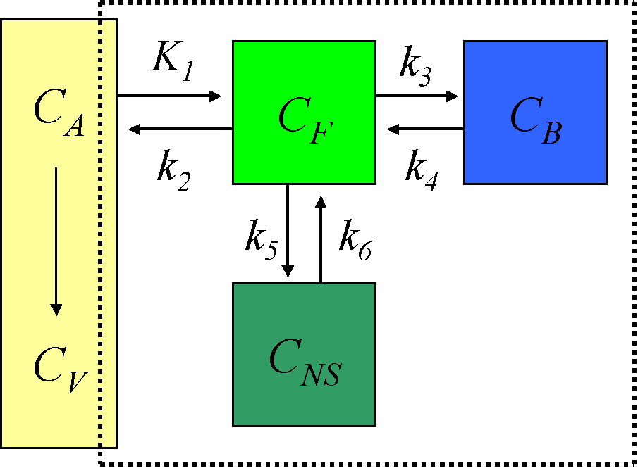
Thus, in principle, the reference tissue should have only one tissue compartment, and the reference tissue curve should follow two-compartmental kinetics.
However, when both models are fitted, the results usually support the three-compartment model. Possible explanations for this are:
Heterogeneity of tissue (mixing of signal from white and grey matter) is certainly always present in PET studies, and can explain the apparent second tissue compartment. The other explanations can contribute to the finding.
Because of these errors, PET measurements can not be used to validate the existence or non-existence of receptors in a suggested reference region. Only blocking or displacement studies can provide definite answers.
Apparent second tissue compartment in reference tissue, if "binding" is low, does not invalidate the use of reference tissue models. If apparent binding is caused by tissue heterogeneity, neither 2- or 3-compartment model is (strictly speaking) correct. The one that provides more accurate results in receptor-containing regions of interest, should be used.
Arterial cannulation should be avoided, when reference tissue methods are applicable. Reference tissue methods also avoid the errors in the determination of the fraction of parent compound in plasma with HPLC or TLC. These errors may easily affect the accuracy of results more than the statistical quality of the PET scans.
However, when a new tracer is introduced and the model and analysis method for it are being developed and validated, the plasma curve and metabolite fractions must be measured.
When metabolism rate is studied no reference tissue usually exists. Thus metabolite corrected plasma data is required. Compartmental models can possibly be applied if carefully validated. For irreversible or nearly irreversible systems Gjedde-Patlak plot can be used [Patlak and Blasberg 1985].
Lammertsma AA. Radioligand studies: imaging and quantitative analysis. Eur Neuropsychopharmacol 2002; 12: 513-516.
Oikonen V, Allonen T, Någren K, Kajander J, Hietala J. Quantification of [carbonyl-11C]WAY-100635 binding: considerations on the cerebellum. Nucl Med Biol 2000; 27: 483-486.
Passchier J, Gee A, Willemsen A, Vaalburg W, van Waarde A. Measuring drug-related receptor occupancy with positron emission tomography. Methods 2000; 27: 278-286.
Patlak CS, Blasberg RG. Graphical evaluation of blood-to-brain transfer constants from multiple-time uptake data. Generalizations. J Cereb Blood Flow Metab. 1985; 5:584-590.
Sawada Y, Hiraga S, Francis B, Patlak C, Pettigrew K, Ito K, Owens E, Gibson R, Reba R, Eckelman W, Larson S, Blasberg RG. Kinetic analysis of 3-quinuclidinyl 4-[125I]iodobenzilate transport and specific binding to muscarinic acetylcholine receptor in rat brain in vivo: implications for human studies. J Cereb Blood Flow Metab 1990; 10:781-807.
Wong DF, Gjedde A, Wagner Jr HN. Quantification of neuroreceptors in the living human brain. I. Irreversible binding of ligands. J Cereb Blood Flow Metab 1986; 6:137-146.