Determination of arterial input function from dynamic PET image
[15O]H2O studies of abdominal region
Arterial blood time-activity curve (TAC) can be estimated from the abdominal aorta in the dynamic PET image. A blood TAC is extracted by thresholding and a recovery coefficient is calculted with a method based on one by Germano et al. For the determination of recovery coefficient, one needs to know the FWHM in the middle of the image (dependent on the scanner AND reconstruction method).
Program for this purpose: eabaort
For example:
eabaort -F=6.3 s06678dy1_mrp.img 10 15 s06678ab.dat
The user needs to check the planes where single aorta is clearly seen
(e.g. ecat2tif).
ECAT 6.3, ECAT 7 formats are supported. Functioning of Analyze format is not
quaranteed. At least that format requires that the frame information must be
added to the result blood file separately
( dftframe).
Output file (.dat) contains the extracted blood curve, which is calibrated and
corrected for decay (see instructions below).
NOTE! Do not name the output file with extension .bld, .lis or .alg.
These are reserved for uncorrected blood files.
Studies with PET-CT
If a CT image is available for the study, it should be used to asses the diameter of aorta. For example in Vinci you can use measure tool from tool box (see figure1). Measure diameter inside the artery wall, DO NOT include the walls. Make sure that the diameter is not varying too much between planes and write down the number of the plane which was used in the measuring. After that, call program eabaort with option -d
eabaort -d=22 -F=6.3 s06678dy1_mrp.img 11 14 s06678ab.datYou still need to know the FWHM value. When you give the plane numbers, you should include the one where diameter was measured and choose planes around it.
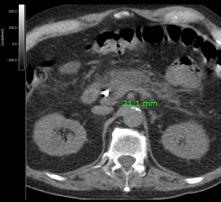 Figure 1. (Click to enlarge)
[15O]H2O studies of femoral region
Same methods will be tested for femoral region in near future.
Usage in calculation of perfusion image
The resulting blood TAC is automatically corrected for physical decay and dispersion, because it is retrieved from the same image as the target tissue. Time delay correction may be still needed (fitdelay). After that the blood curve can be used directly from step #2 to calculate perfusion image using autoradiography.
However, if old SUN program FlowImg is used in autoradiography, the resulting blood file must be interpolated to have 1 sec sample times, as required by this program. Also, it is useful to change the filename to have extension .fit.
For example to interpolate the TAC from 0 to 300 sec:
interpol -a -c0,300,1 s06678ab.dat s06678ab.fit
The interpolation time should be set from 0 to the end of last PET frame, but at least to the end of integration time.
Instead of autoradiography, also compartment model fitting can be used to produce perfusion images with program imgflow. This program can read directly the blood TAC produced with eabaort.
