Compartmental models
If metabolite corrected arterial blood curve and dynamic PET data are available from the time of injection to the time where all important changes in tracer kinetics have been seen, and the data is of sufficient quality, then it may be possible to estimate the parameters of a complete model, including perfusion, blood volume in tissue vasculature, transport, specific binding or reaction rate etc. In practice this can be done only in very simple cases, e.g. with labeled water which just flushes in and out of tissue. As is mentioned above, it is possible and desirable to either measure some of the parameters in separate studies, e.g. blood volume using [15O]CO, or constrain them to literature values, or to reduce the model. Most often there is no need to measure all the parameters, but just one key parameter which correlates with the desired property in usual conditions.
Plasma compartment
To be precise, the plasma is not a compartment of the model. The concentration of tracer in the plasma is measured, and applied to the compartment model as a known input function. However, plasma compartment is still usually counted as one of the compartments.
The metabolite corrected arterial plasma curve is the input to the compartment model. If the intravascular activity is accounted for in the calculation, the whole blood concentration, containing metabolites, should be used for that. If metabolite corrected plasma curve is used instead of uncorrected whole blood curve to correct for vascular blood volume fraction, blood contribution at late times is underestimated, which could result in the artificial presence of an apparent additional tissue compartment [Lammertsma 2002].
Two-compartment model (one-tissue compartment model)
Methods for quantitation of perfusion (blood flow, f) withfreelydiffusable tracers are based on Kety's analyses of the principles of inert gas exchange. Tracers such as [15O]H2O, [15O]butanol, [11C]butanol and [18F]fluoromethane are used for this purpose. Also a single breathinhalation of [15O]CO2 produces an arterial bolus of [15O]H2O.
Three-compartment model (two-tissue compartment model)
The kinetic model for measurement of glucose transport and phosphorylationrate in brain is based on a three-compartment model.
Four-compartment model (three-tissue compartment model)
The description of the time course of the ligand in tissue rquires a modelthat accounts for the different components contributing to the signal. These are free ligand in plasma, free ligand in tissue, CF, ligand in tissue which is not specifically bound, CNS, and ligand specifically bound to the receptor, CB [Schmidt and Turkheimer 2002]:
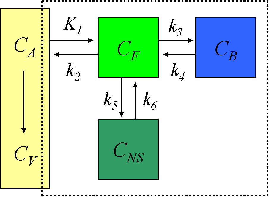
Use of 4-compartment model is not feasible given the large number of rate constants to be estimated, and the kinetically undistinguishable compartments for specific and non-specific binding. The model is simplified under the assumption of a rapid equilibrium between free and non-specifically bound compartments that produces a single compartment of free + non-specifically bound ligand:
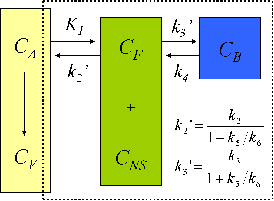
Although for most ligands k3' and k4 cannot be estimated with a reasonable degree of accuracy, their ratio is usually more stable.
