Reference tissue compartmental models
Reference tissue (input) compartmental models do not require or use plasma samples, but instead are using time-activity curve of a reference region with non-existent (or very low) specific binding. Usually reference tissue models are used to estimate binding potential (BPND) from reversible ligand-receptor PET studies, but there are also modified models that can be applied to irreversible binding.
Full reference tissue compartment model
The (full) reference tissue compartmental model [Cunningham et al 1991] is the original and "gold standard" of reference tissue input methods for the estimation of binding potential (BPND) from reversible ligand-receptor studies. In most cases, it can be replaced by the simplified reference tissue model.
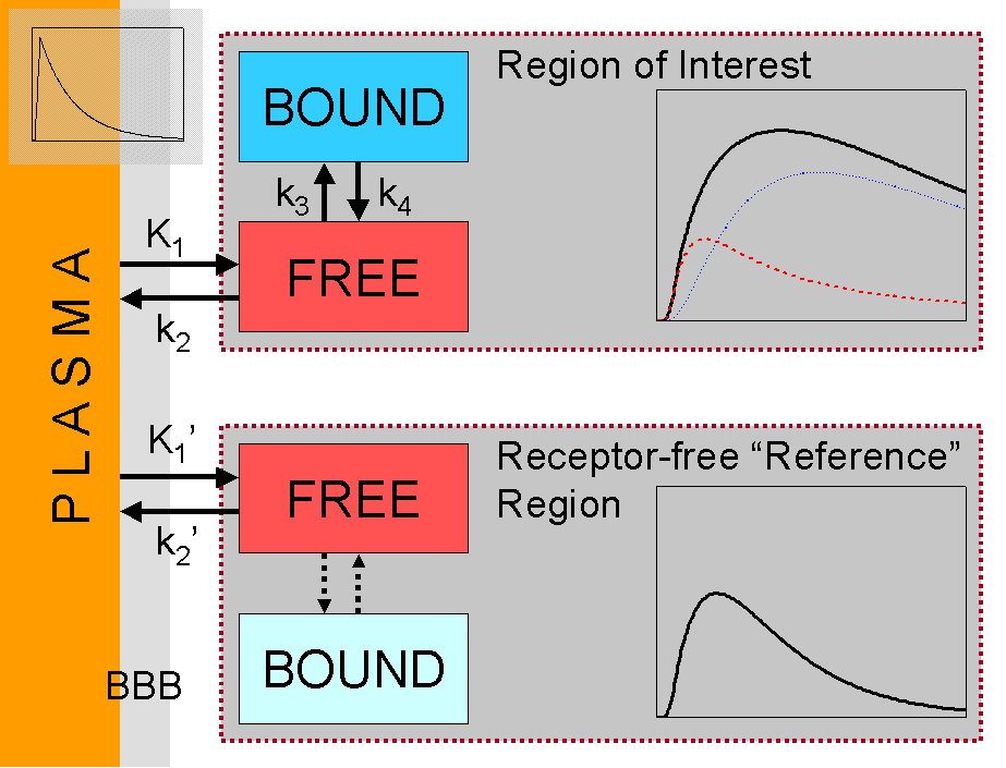
The parameters R1 (ratio of the K1 values of regions of interest and reference tissue), k2, k3, and BPND (k3/k4) can be estimated using nonlinear fitting [Cunningham et al 1991]. This model has some advantages over the Logan plot: dynamic study can be used from the beginning with no need to wait for any equilibrium or search for linear phase. Reference tissue model also provides an estimate for the perfusion and transport of tracer to the tissue (R1).
If the binding is irreversible, the model can be reduced by setting k4=0. Then the k3 value provided by the model will be proportional to the concentration of unoccupied receptors [Wong et al. 1986].
Simplified reference tissue model (SRTM)
Simplified reference tissue (or region) model (SRTM or SRRM) can be used when two compartmental model could reasonable describe the kinetics of the tracer in tissue (Lammertsma and Hume 1996). The parameters of simplified model (R1, k2 and BPND) can be solved not only using nonlinear fitting but also using linearized methods (Blomqvist 1984), or with basis function approach (Gunn et al. 1997). This makes it possible to produce parametric images of model parameters, and, when linearized, also to do the calculations at the sinogram level.
Basis function implementations
Basis function implementation of SRTM (RPM, Gunn et al., 1997) produces unbiased BP maps presuming that the range of basis functions is carefully optimized. In practice, selection of a specific range of basis functions is slow and inefficient and is based on a compromise between accuracy and precision (Cselényi et al., 2006; Schuitemaker et al., 2007).
Wu and Carson (2002) proposed that the washout rate constant of the reference tissue could be first estimated using RPM, and the median value (including voxels where BP>0) would be fixed for all voxels during the second basis function evaluation. This approach mostly improves the quality of R1 images, but leads to negative bias in low BP values (Schuitemaker et al., 2007).
Modified SRTM to detect neurotransmitter release
SRTM assumes a steady physiological state throughout the PET experiment, from radiotracer injection to the end of scanning. Steady-state can be intentionally perturbed to study the effect of task- or drug-induced changes in neurotransmission (Friston et al., 1997). To analyze this kind of data the reference tissue model had to be modified to account for the changes in the dissociation rate of the radioligand (LSSRM, Alpert et al., 2003).
Ichise's multilinear regression techniques
Ichise et al (1996) presented a method in which the equations of Logan graphical analysis are solved with multilinear regression. This approach may lead to marked negative bias with noisy data (Schuitemaker et al., 2007). The modified method (Ichise et al., 2002 and 2003) may lead to higher variance withslightly reduced bias (Schuitemaker et al., 2007).
