Blood and plasma TACs
All quantitative analysis methods require that an arterial input function is measured, or an appropriate surrogate is found.
Manual samples are measured with automatic gamma counter (Wizard 1480 3", Wallac, Turku) in the "blood laboratory" located besides the PET scanners. When necessary, plasma is separated by centrifuging, and then measured.
By default, the radioactivity concentrations in manual samples of blood or plasma are corrected for physical decay to the injection time, and calibrated to units kBq/mL. Laboratory personnel copies the plasma and/or blood TAC datafiles to the PETO system. From there, the datafiles can be copied to your group disks.
Datafiles are in ASCII format (File format of time-radioactivity curves), that can be imported e.g. to Excel, Origin or OpenOffice Calc. See also plotting of TACs.
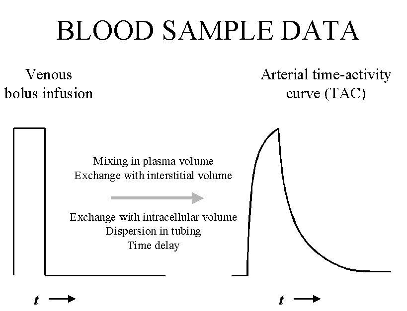
Delay and dispersion
Arterial curve is dispersed in the sample tubing and the arrival times to the region of interest and to the sampling point may be different. Thus the input curve may need to be corrected for time delay and dispersion.
Fitting plasma curves
Fitting mathematical function to plasma curve.
Averaging several measured plasma curves.
Extrapolate plasma curves with1-exponential function
Pharmacokinetics
Calculating pharmacokinetics from plasma curve.
